Clinical AI · Chest Radiography · Computational Biology

Artificial intelligence can make routine chest X-rays more useful at the very first visit.

A curated overview of how AI is used to analyze chest radiographs and MRI, work with DICOM studies, support first-line screening in ordinary clinics and design new therapeutic proteins and antibodies.

Educational information only. Demonstration figures are for illustration and do not provide diagnosis or treatment.
Multimodal clinical AIresearch
X-ray / MRIDICOMScreeningBiologics

AI + chest radiography

Scalable screening for ordinary outpatient clinics

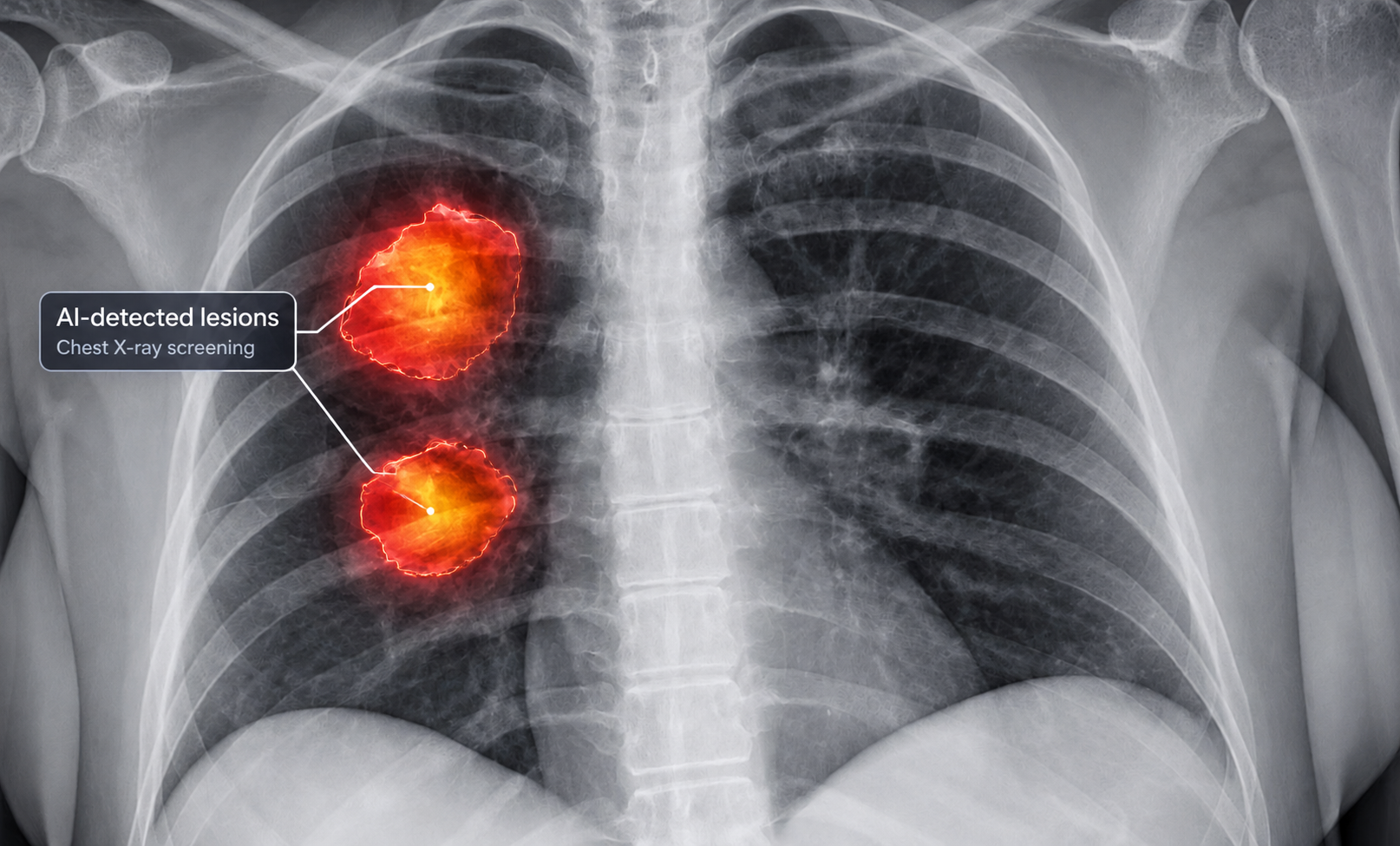
Chest X-ray is cheaper, faster and far more widely available than CT. That makes it attractive for mass checkups, occupational screening, routine dispensary programs and a first visit in ordinary polyclinics. AI can help by flagging suspicious findings on plain radiographs so that patients who need closer review or CT can be prioritized earlier.

Chest X-ray with AI-highlighted suspicious lesions
Chest X-ray · Triage

AI-assisted lesion detection on radiography

On a standard chest radiograph, subtle nodules or focal opacities can be easy to miss, especially in busy first-line care. AI systems can scan the image, highlight suspicious regions and support a faster referral pathway for radiologist review, repeat imaging or CT confirmation.

Illustrative demonstration figure for educational use.

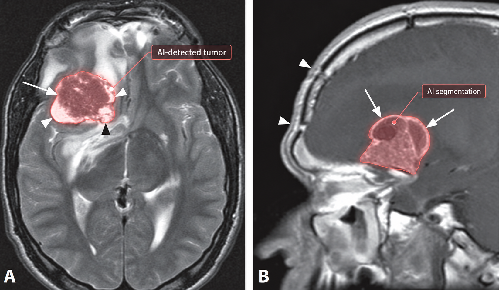
Brain MRI with AI segmentation overlay for a tumor
MRI · Segmentation

AI also helps with more advanced imaging

For MRI, segmentation models can outline tumor boundaries and enable volume tracking over time. This is a different use case from population screening, but it shows how AI ranges from simple frontline triage on X-rays to detailed quantitative support on complex scans.

Illustrative demonstration figure for educational use.

AccessibleChest X-ray is available in most routine clinics.
Low costUseful where CT cannot be used for everyone.
TriageAI can surface studies that deserve closer human review.
EscalationSuspicious cases can be referred for CT and specialist workup.
A 2025 review in npj Precision Oncology describes AI applications across lung cancer prevention, screening, diagnosis and treatment, including the use of chest X-rays and routine clinical data for risk detection. Read the review ↗

DICOM in practice

A CT study is more than a stack of JPEGs.

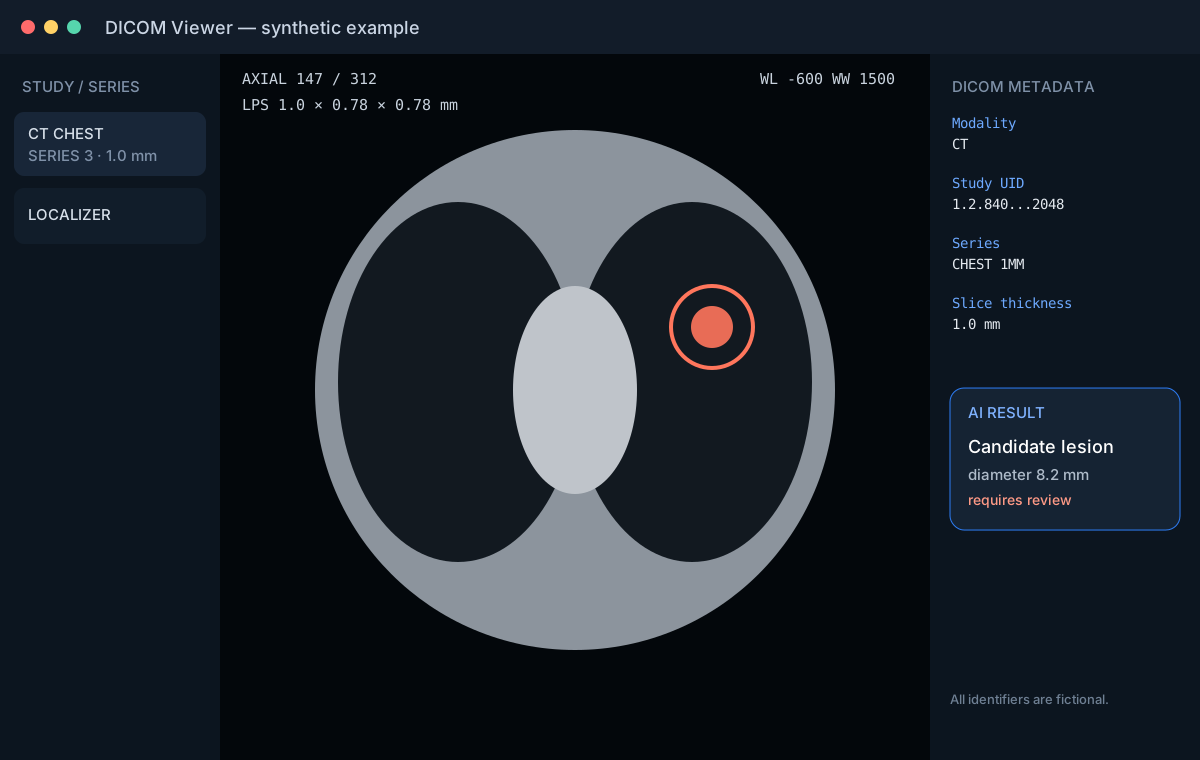
DICOM carries pixel data together with structured metadata that identifies the study, series, modality, acquisition parameters and spatial context. That matters not only for CT and MRI, but also for digital radiography workflows when AI results need to return to PACS cleanly and consistently.

Read the current DICOM PS3.18 standard ↗
Synthetic DICOM viewer with CT series, metadata and AI overlay
Illustrative viewer. Example identifiers are fictional.
01Study arrives

A chest X-ray, CT or MRI study is stored in PACS/VNA with DICOM metadata.

02AI preprocessing

Series selection, resampling, normalization and quality checks.

03Inference

The model produces detections, masks, measurements or probabilities.

04Clinical review

Results return to the workflow as overlays, structured data or triage signals.

Generative AI + Biologics

AI can search protein sequence and structure space, not just pixels.

For therapeutic antibodies, computational models are being used for structure prediction, epitope targeting, affinity optimization, developability screening and de novo generation. Experimental validation is still essential: a model proposal is not a drug.

Y
Research milestone

De novo antibody design with RFdiffusion

A 2025 Nature study reported computational generation of antibody formats aimed at user-specified epitopes, followed by experimental screening and structural characterization. It is a concrete example of generative protein design moving beyond sequence ranking.

Nature paper ↗
HER2
Industry example

Absci: AI-designed anti-HER2 program

Absci has publicly described ABS-501 as an AI-designed anti-HER2 antibody program. The company reports preclinical leads with novel epitope interactions and activity in a trastuzumab-resistant xenograft model. These are company-reported preclinical data, not evidence of clinical efficacy.

Company update ↗
ADC
Oncology review

AI across the antibody-drug conjugate pipeline

A 2025 review in npj Precision Oncology describes AI applications spanning tumor-target selection, antibody engineering, linker/payload optimization, ADMET modelling and patient stratification.

Open-access review ↗
Important distinction: AI can generate or rank candidate molecules, but therapeutic development still requires biochemical assays, cell and animal studies, manufacturing assessment, toxicology, clinical trials and regulatory review. “AI-designed” does not mean “clinically proven.”
Responsible clinical AI

A strong demo is not the same as a safe screening program.

External validation

Performance should be tested beyond the development dataset, ideally across sites, scanners and patient populations relevant to intended use.

Calibration & failure modes

A model can have high discrimination and still produce poorly calibrated probabilities or systematic errors in important subgroups.

Workflow design

The way an alert is shown, escalated and documented matters, especially when AI is used at scale in routine outpatient screening rather than in a single specialist center.

About this project

Independent educational resource

AI in Medicine collects examples, papers and technical concepts at the intersection of medicine and artificial intelligence. It does not provide medical advice, diagnosis or treatment.

contact@aiinmedecine.top